Blog
Age-Related Macular Degeneration
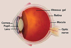
What is AMD?
AMD is damage to the macula of the eye in people age 50 and older. The macula is the central part of your retina. It is made up of millions of light-sensing cells that are responsible for sharp central vision. The retina quickly turns light into electrical signals and then sends them to your brain through the optic nerve. Your brain translates the electrical signals into the images you see.
If your macula becomes damaged, fine points in these images are no longer clear. The overall picture might still be there, but the fine points are missing. Some people with AMD view straight lines as “squiggly.” Others see a blind spot in their central vision. AMD does not lead to complete blindness, but it can interfere with your ability to do daily activities like reading, driving, writing, or cooking.
What causes AMD?
Age is the leading cause of AMD, but these factors can also raise your risk:
- Smoking, which doubles AMD risk
- Race. AMD is more common among whites than African-Americans or Hispanics.
- Genetics. AMD has been linked with certain genes and can run in families.
- Obesity. Being overweight increases your risk of developing AMD.
What is the difference between wet and dry AMD?
In people with “wet” AMD, abnormal blood vessels behind the retina start to grow under the macula. These new blood vessels can be fragile, leaking blood and fluid that cause the macula to swell. Damage occurs rapidly and may also cause scarring of the retina. About 10 percent of people with AMD have wet AMD. “Dry” AMD happens when the light-sensitive cells in the macula slowly break down. As dry AMD progresses, patients may see a blurred spot in the center of their vision.
How would I know if I had AMD?
The best way to learn if you have AMD is to get regular eye exams from an eye care professional, including a vision test and dilated eye exam. Your doctor will look for drusen (yellow deposits) or pigment changes beneath your retina. Other tests used to diagnose AMD include:
- Amsler grid. Your doctor may ask you to look at an Amsler grid, which is a simple grid of black lines on a white background. If you have AMD, the lines of the grid may disappear (especially in the middle) or appear wavy.
- Fluorescein angiogram. A fluorescent dye is used to see the blood vessels in your eyes to determine if they are leaking.
- Optical coherence tomography is an imaging test that uses light waves to capture images of your eye tissue.
Some people with vision loss have visual hallucinations called Charles Bonnet syndrome. The brain loses input from the eyes and fills the void by generating its own visual images that are not really there. Charles Bonnet syndrome usually lasts a year to 18 months after it starts, and the hallucinations happen more frequently in dim light or in the evening. Tell your doctor if you are having visual hallucinations.
How is AMD treated?
Your treatment for AMD depends on the type and stage of the disease. Early AMD typically requires no treatment; your doctor will want to see you at least once a year for a dilated eye exam and may recommend that you exercise regularly, eat a healthy diet rich in leafy green vegetables and fish, and avoid smoking to slow the progression of AMD.
If you have intermediate AMD, your doctor may recommend certain vitamin supplements to slow disease progression, These include high doses of vitamins C and E, zinc, copper, lutein, and zeaxanthin. Do not take these supplements on your own; see your eye doctor to determine if they are recommended for you and at what doses.
Advanced wet AMD results in severe vision loss. The goal of treatment is to stop the growth of new blood vessels and the swelling that occurs in wet AMD.
- Injections. The protein “vascular endothelial growth factor” (VEGF) is overproduced in the eyes of people with wet AMD. Therapies that target VEGF—such aflibercept or ranibizumab—inhibit VEGF activity and blood vessel growth. These treatments are injected into the eye. In some patients, anti-VEGF therapies can improve vision, while in others, the goal is to prevent wet AMD from getting worse.
- Photodynamic therapy. A drug called verteporfin is injected into a vein and is absorbed by new, growing blood vessels. When the doctor shines a “cold” laser beam into the affected eye, the drug is activated to close off the new blood vessels, slowing their growth and the rate of vision loss.
- Laser surgery. Though not used as often as other treatments, this form of AMD therapy employs a hot laser to destroy abnormal blood vessels that are limited to a compact area of an eye, away from the center of the macula.
- Research continues to find better treatments for AMD. One approach is to make stem cells from a patient’s own skin or blood and grow them to form the pigmented layer of tissues that supports the retina.
How can I learn to live with AMD?
AMD causes low vision. Even with regular glasses, contact lenses, medications, or surgery, you may struggle with everyday tasks. Talk with your eye care professional about your vision problems and ask about vision rehabilitation: guidance about things you can do to cope with your AMD and maximize your remaining vision. Low vision services are available in certain ophthalmology and optometry offices, hospital clinics, independent living centers, and vision rehabilitation organizations and may include:
- Reading glasses with high-powered lenses
- Large-print reading materials
- Computer aids and other technologies
- Handheld magnifiers
- Talking devices such as watches, calculators, and clocks
- Video magnifiers
- Implantable miniature telescope (IMT), a surgically implanted device for end-stage AMD designed to refocus some images onto a healthier part of the retina
Adjusting to life with AMD can be challenging, frustrating, and depressing. Build a support team to help you learn to live with AMD, and try to educate yourself about vision loss. Stay engaged with family and friends, ask for help when you need it, and consider seeking a professional counselor or support group.
Source: The National Eye Institute (NEI)